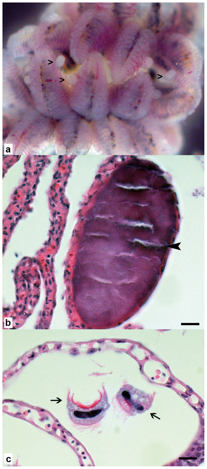
Figure 1. Wet mount and histology of pipefish gills.

a: Wet mount in sterile sea water with numerous protruding epitheliocystis lesions clearly visible (open arrowheads). b: Gill lamella with focal intracellular cyst in epithelial cell, 40 µm long axis, with dark basophilic granular material (black arrowhead), the epithelium of affected lamella shows no pathological changes and no inflammatory reaction, scale bar = 10 µm. c: multiple Trichodina sp. between lamellae (arrows), no associated pathological changes, scale bar = 10 µm.
