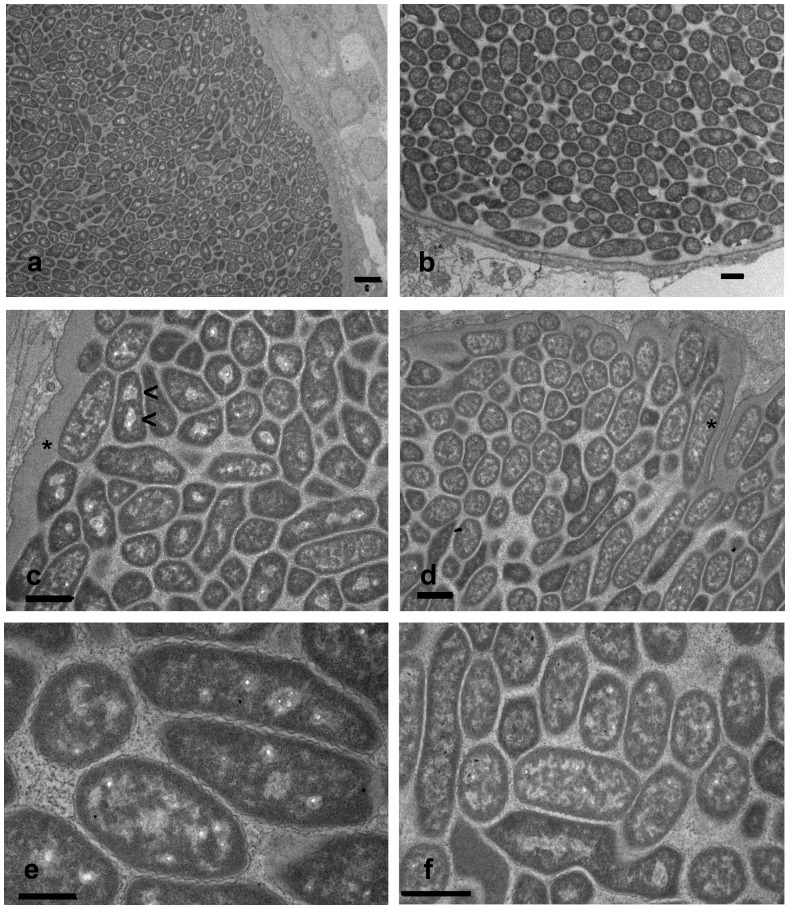
Figure 4. Transmission electron microscopy of epitheliocystis lesions showing features typical for Candidatus Syngnamydia venezia.
Both a and b give an overview of the dense cell packing. In c and d, endosymbionts showing one or two electron-lucent regions (<), possibly representative of dividing bacteria, and maximally 3.4 µm (d, *) in length. The rippled outer membrane (e) as well as the angular forms (f) are typical characteristics. Scale bars = 1 µm.

