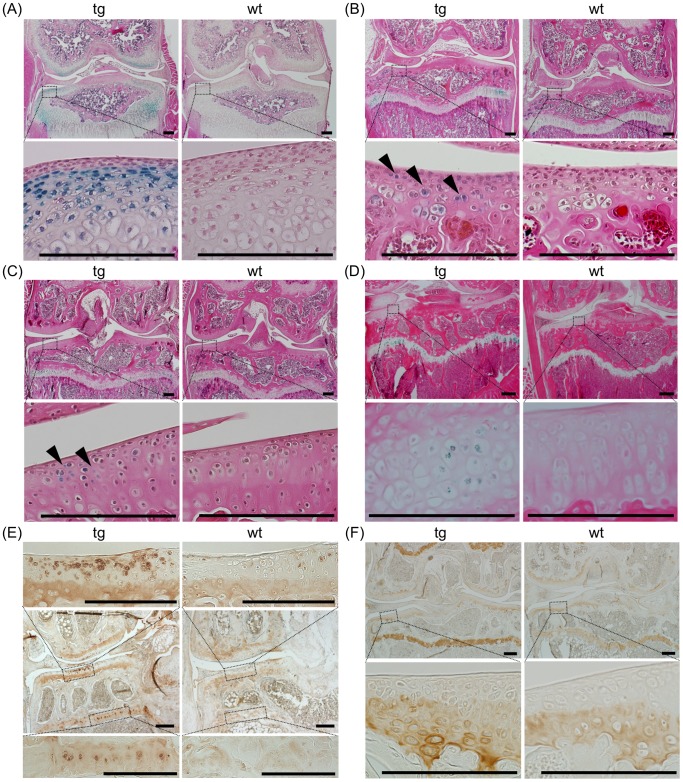
Figure 1. Overexpression of CCN2 and accumulation of type II collagen in aged articular cartilage of transgenic mice.
(A–D) Frontal sections of knee joints after X-gal staining. Specimens from 14- (A), 40- (B), 60- (C), and 150-day-old (D) TG (left) and WT (right) mice. Lower panels show magnified regions of tibial articular cartilage as indicated by the dotted boxes in the upper panels. Expression of LacZ in the articular cartilage was detected in the TG sections. Arrowheads indicate LacZ-positive articular chondrocytes. Bars: 200 µm. (E) Immunohistochemical staining of CCN2 in frontal sections of knee joints from 21-month-old TG and WT mice. Upper and lower panels show magnified load-bearing regions of tibial articular cartilage (upper) and growth-plate cartilage (lower), as indicated by the dotted boxes in the middle panels. Bars: 200 µm. Deposition of CCN2 in the upper zone of TG articular cartilage was enhanced compared with that in WT articular cartilage. (F) Immunohistochemical staining of type II collagen in frontal sections of knee joint from 21-month-old TG and WT mice. Lower panels show magnified load-bearing regions of tibial articular cartilage, as indicated by the dotted boxes in the upper panels. Bars: 200 µm. Accumulation of type II collagen is enhanced in TG growth-plate cartilage as compared with WT, but in articular cartilage the difference was not significant.

