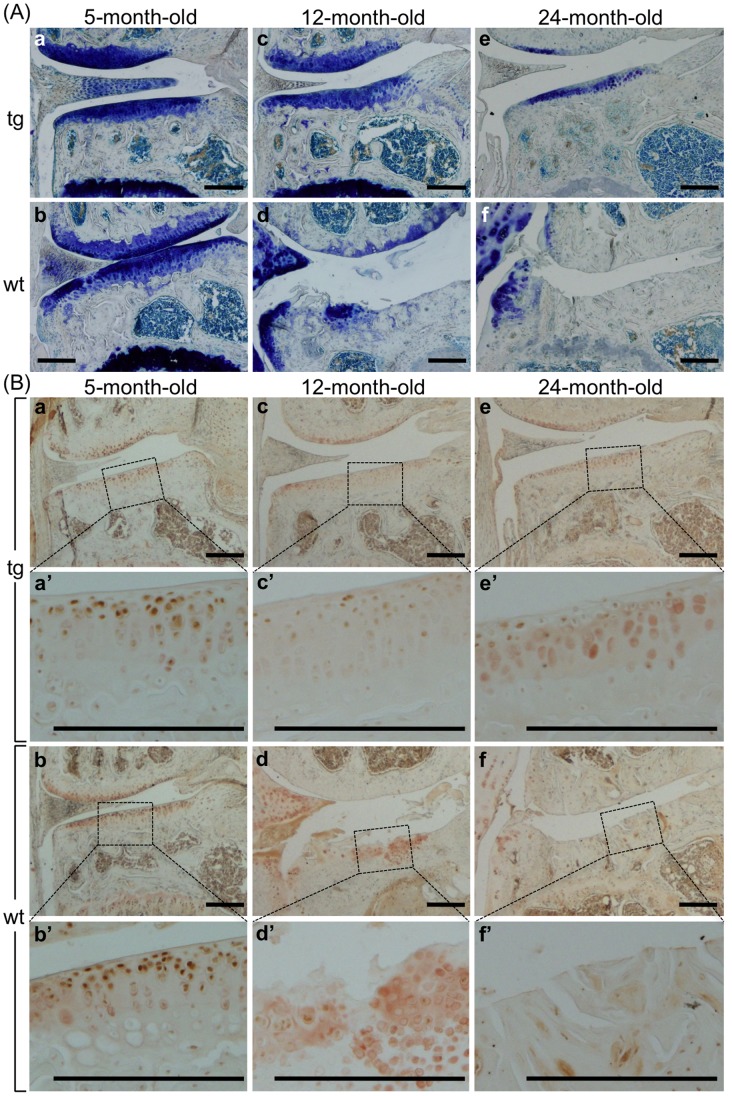
Figure 7. Preventing age-related degeneration of proteoglycans in articular cartilage by CCN2 overexpression.
(A) Toluidine blue staining of frontal sections of medial portion of TG (a, c, e) and WT (b, d, f) littermate knee joints from 5- (a and b), 12- (c and d), and 24- (e and f) month-old male mice. TB staining indicated that WT articular cartilage was degraded with age, and finally the whole layer was lost after 24 months. (B) Immunohistochemical staining of aggrecan neoepitope in frontal sections of the medial portion of TG (a, c, e) and WT (b, d, f) littermate knee joints from 5- (a and b), 12- (c and d), and 24-month-old (e and f) mice. a’, b’, c’, d’, e’, and f’: higher magnification of the medial tibial plateau in the load-bearing region, indicated by the dotted box in a, b, c, d, e, and f, respectively. Bars: 200 µm. In the WT tibia (d and d’) even the remaining matrices were slightly degraded, as shown by the aggrecan neoepitope staining. TG cartilage showed a minor decrease in staining for the aggrecan neoepitope (a’, c’, and e’), but not severe as in the WT cartilage (b’, d’, and f’).

