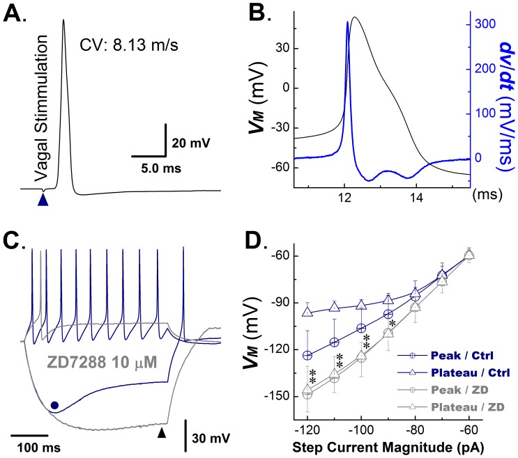
Figure 3. Ih in myelinated Ah-type VGNs from adult, non-ovariectomized rats.
A) Vagal stimulation-evoked transmembrane action potential in a myelinated Ah-type vagal ganglion neuron (VGN). The value for the conduction velocity (CV) measured between the stimulation and recording site was indicative of an Ah-type cell. B) Transmembrane action potential recorded from the same neuron as in (A) and its first derivative over time (blue trace). Note the presence of a repolarization ‘hump’. C) Examples of membrane potential responses of an Ah-type VGN to a depolarizing and a hyperpolarizing current injection step. The cell was first injected with 150 pA current step, giving rise to repetitive action potential firing, which ceased upon termination of the current injection (blue trace). A −120 pA step current injection caused membrane hyperpolarization (blue trace), which gradually increased to its maximum value (−130 mV, dot in Figure 4C) and then depolarized slowly (−96 mV, sag) despite continued current injection. Return of the membrane potential to baseline was associated with the occurrence of a spontaneous action potential. Gray traces show the membrane potential changes in response to a positive and negative current step injection in the presence of the Ih blocker ZD7288 (10 microM/L). ZD7288 suppressed spontaneous action potential discharge during depolarizing current injection, and reduced the sag potential (difference between peak membrane potential and endpulse potential). D) Plots of the peak vs. endpulse voltage as a function of the magnitude of the hyperpolarizing step current injection under control and following application of ZD7288. ZD7288 suppressed sag potentials in Ah-type neurons. Date are mean ±1 SD. n = 6 cells for each data point, *P<0.05 and **P<0.01 vs. control (Ctrl).

