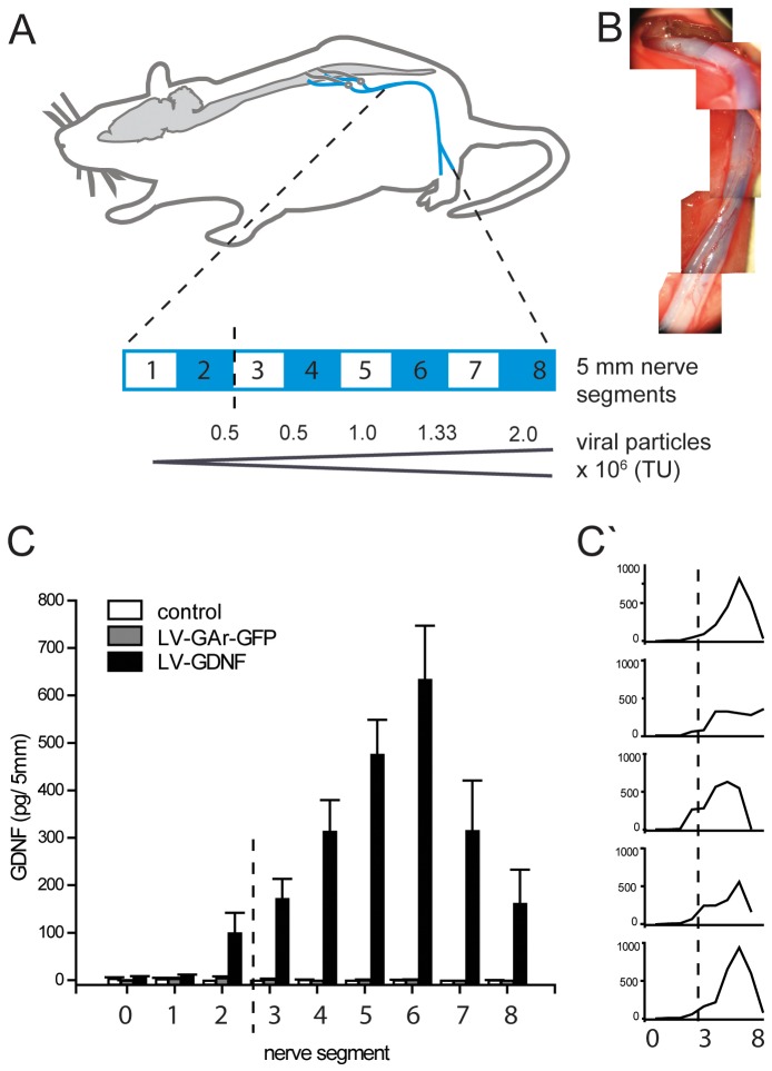
Figure 2. Creating a GDNF gradient in the intact rat sciatic nerve by injecting increasing amounts of LV-GDNF along the nerve.
A) Overview of the spinal cord, lumbar ventral roots and the sciatic nerve with a schematic enlargement of the nerve that was injected with increasing amounts of LV-GDNF ((0.25–2×10⁶ TU) or LV-GArGFP at 5 locations at 7 to 8 mm intervals. After four weeks, the sciatic nerve is removed and divided in 5 mm segments for GDNF quantification. The blue-white bar depicts the position of each harvested segment, the dotted line marks the epineural suture visible in (B). The approximate vector injection site is shown below the blue-white bar. B) Per-operative photographs of the sciatic nerve after injection of LV-GDNF mixed with Fast green (a dye) to visualize the injection. An epineural suture, marking the most proximal injection, is visible in the proximal end of the nerve. Note the even spread of the viral vector solution along the entire length of the nerve. C) Total GDNF protein in homogenized 5 mm nerve segments, 4 weeks after injection. A clear gradient of increasing GDNF levels is present in the LV-GDNF group, but not in the LV-GArGFP or non-injected control groups. The dotted line corresponds to the site of the epineural suture visible in (B). C’) GDNF protein levels in individual animals: although there is a slight variation in the location and level of GDNF expression, a gradient is present in the nerve of each animal.

