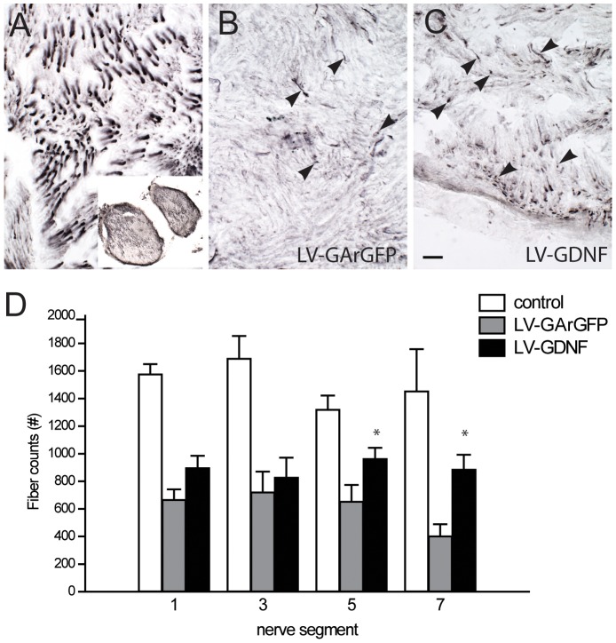
Figure 6. The LV-GDNF gradient leads to significantly more motor axons in the distal sciatic nerve 17 weeks after ventral root avulsion and reimplantation.
ABC) ChAT staining of transverse sections of a non-avulsed sciatic nerve (a) shows thick motor axons within their nerve fascicles. Inset: overview of transverse area of the entire nerve. In avulsed and LV-GArGFP (B) or LV-GDNF (B) injected nerves, there are fewer ChAT positive motor axons (arrowheads), they are thinner and lack the longitudinal alignment of unlesioned axons. Scale bar in C: 25 um. D) The number of axons in lesioned animals is lower than intact control nerves. Whereas the number of motor axons declines in the distal segments of the nerve in LV-GArGFP treated animals, the number of axons remains constant after LV-GDNF treatment. In the 5th and 7th segment, the number of fibers is significantly higher after application of the GDNF gradient, *p<0.05 vs LV-GArGFP. Note that coil areas were excluded from fibre quantification.

