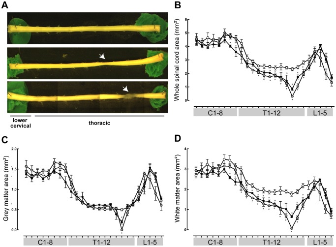
Figure 3. Morphometric measurements of Monodelphis spinal cords.
Control (n = 5, white circles), P7-injured (n = 3, black circles) and P28-injured (n = 4, grey circles) opossum spinal cords. A: Representative whole mounts of lower cervical and thoracic spinal cords from control (top), P7-injured (middle) and P28-injured (bottom) Monodelphis. B: Whole spinal cord cross-sectional area along the length of the spinal cord. C: Grey matter area. D: White matter area. Mean ± sem. *P≤0.05 vs control. Stars denoting significance appear immediately above data for P7-injured animals or immediately below data for P28-injured animals.

