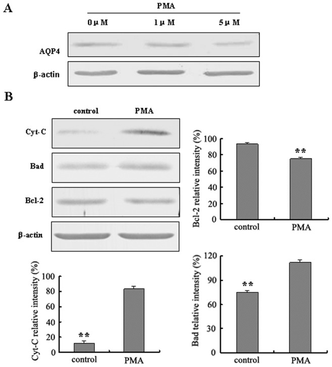
Figure 6. Reduction of AQP4 by PMA in LN229 cells induced apoptosis.
A. The expression of AQP4 in LN229 cells with PMA (0 µM, 1 µM, 5 µM) treatment (24 hr) was detected by Western blotting. B. Western blotting was performed by using anti-Cyt-C, anti-Bad or anti-Bcl-2 antibody. (5 µM of PMA was used.) Expression of Cyt-C, Bad and Bcl-2 were quantitated by densitometry and normalized to β-actin expression. Data were analyzed using two-way ANOVA analysis **P<0.01.

