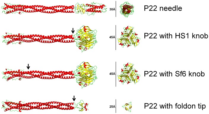
Figure 3. Structural models of the P22 gp26 tail needle and chimeric tail needles.
A. Crystal structure of P22 tail needle gp26 (pdb 3C9I). B–D. Homology structural models of chimeric P22 needles with Sf6 knob (in phage UC-0911), HS1 knob (in phage UC-0926) and foldon tip (in phage UC-0927). Chimeric models were generated for illustration with align function of PyMol (Version 1.3, Schrodinger, LLC, San Carlos, CA), where C-terminal knob domains of Sf6 and HS1 tail needle (pdb 3RWN, 4K6B), C-terminal foldon domain from fibritin fiber of the bacteriophage T4 (pdb 1AA0) were fused downstream of P22 gp26 tail needle helical core residues 1–140 (pdb 3C9I), respectively. In all three models, arrow indicates point of fusion.

