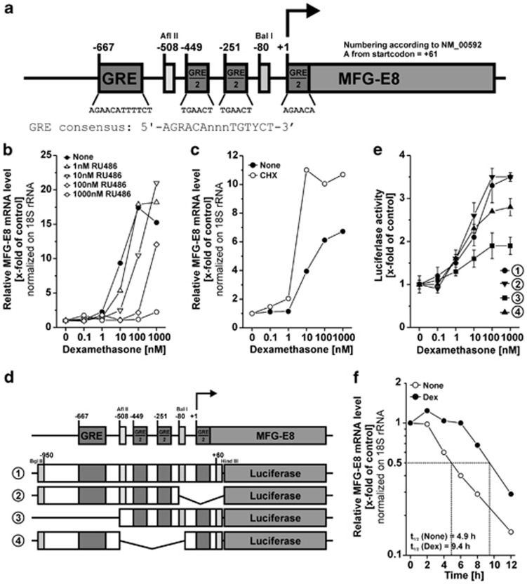
Figure 3.
Different GREs in the promoter region of human MFG-E8 are required for dexamethasone-dependent MFG-E8 expression. (a) Schematic map of putative GREs within the minus 1000-bp region of the human MFG-E8 promoter. Positions of GREs and restriction sites are numbered according to NM_00592. (b) THP-1 macrophages were treated with Dex in the presence or absence of 10 μM RU486 for 24 h. MFG-E8 mRNA levels were quantified by qRT-PCR as in Figure 1c (means of duplicates from one representative out of two experiments). (c) THP-1 macrophages were treated with Dex in the presence or absence of 10 μg/ml of cycloheximide (CHX) for 24 h. MFG-E8 mRNA levels were assessed as in Figure 1c (mean values of duplicates from one representative out of two experiments). (d) Schematic map of the MFG-E8 promoter constructs employed in (e). (e) Different regions of the human MFG-E8 promoter cloned into pGL3 Basic (see (d)) were lipofected into THP-1 macrophages for 5 h and subsequently cells were treated with Dex for 16 h. Luciferase activities were measured in cellular lysates and calibrated on the corresponding untreated control populations (mean values±S.D. of triplicates from one representative out of three experiments). (f) THP-1 macrophages were treated with or without 1 μM Dex for 2 h before RNA synthesis inhibition with 100 ng/ml actinomycin D. The decay of MFG-E8 mRNA was measured by qRT-PCR. Half-lives (t1/2) of MFG-E8 mRNA were determined and are depicted in the plot (means of duplicates from one representative out of two experiments)

