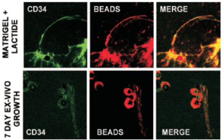
Fig 1.
Matrigel implants were thinly sliced, and cells were labeled with specific anti-mouse CD34-FITC in 1:100 dilutions for 60 min on ice and then placed onto a glass slide for observation. CD34 is the surface marker of SPC. The presence of functional vascular channels in the Matrigel was documented by injecting mice with 40nm carboxylate-modified FluoSpheres® conjugated to Nile red. Top row is SPC surface marker CD34 expression and vascular channels identified by Nile red beads in Matrigel harvested 18 h post implantation. The bottom row shows images of Matrigel incubated ex vivo for 7 days.Note that each row shows images from different samples. Even at just 18 h after implantation, channels could be visualized. CD34 cells lined these channels, indicating vasculogenesisof SPCs in Matrigel. (Reprinted and adapted with permission from ASM, reference 46.)

