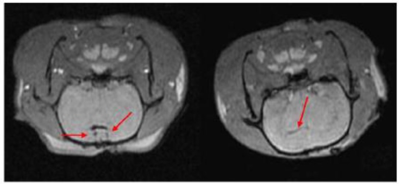
Fig 6.

MRI of transplanted Resovist®-labeled NSCs in the rat brain. At 1 hour following transplantation of Resovist®-labeled NSCs (T2 fast-gradient echo sequence), arrows reveal two symmetrical and horizontal pinholes (left). At 2 weeks post-transplantation, the arrow reveals the migration of Resovist®-labeled NSCs from the right lateral ventricle to the ischemic area (right). (Reprinted and adapted with permission from NRR, reference 87).
