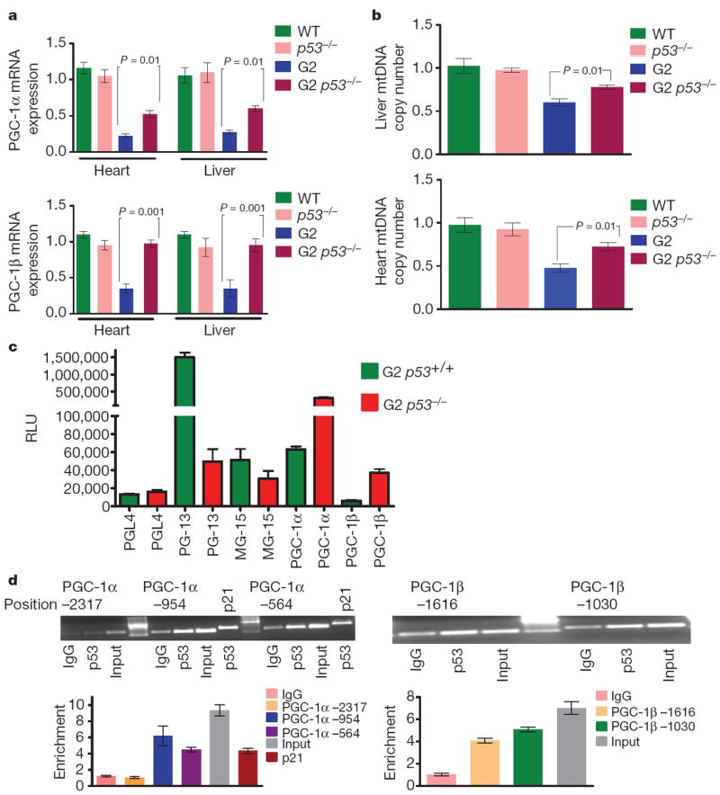
Figure 4. p53 deficiency partially rescues the transcriptional regulation of PGC-1α/β and mitochondrial DNA copy number.

a, PGC-1α and PGC-1β expression in liver and heart (n = 4). b, mtDNA quantification in liver and heart (n = 4). c, p53 represses PGC-1α and PGC-1β promoter reporters. G2 Terc p53+/+ and G2 Terc p53−/− MEFs were transfected with empty reporter pGL4 or PGL4 containing various reporter fragments (−2.8 and −2.6 kb fragment of mouse PGC-1α and PGC-1β). Controls included the PG13-luc plasmid (containing 13 copies of a synthetic p53 DNA binding site) and MG15-luc (containing 15 copies of a mutated p53 DNA binding site). Shown is the average luciferase value (relative light units, RLU) of three different experiments. d, Chromatin immunoprecipitation (ChIP) showing p53 binding on the promoters of PGC-1α and PGC-1β at indicated sites and at p21 site (positive control). Graphs below show quantitative results in the proximal promoter regions by qPCR (three independent experiments). ΔΔCt method was used to analyse RT–qPCR data and t-test was used to calculate the statistical significance, error bars represent s.e.m.
