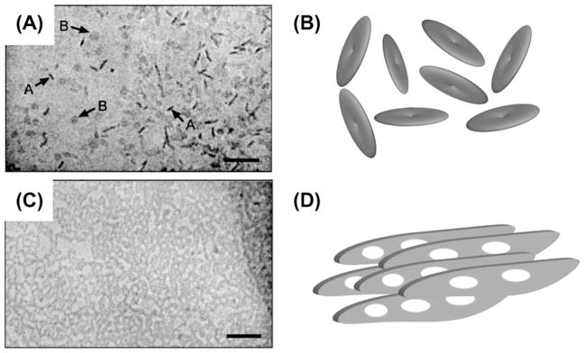
Fig. 1.

Lipid bicelles are supramolecular aggregates that are formed when appropriate amounts of lipids and detergents are mixed in an aqueous environment. The size and phase of bicellar aggregates depend on the [lipid]:[detergent] ratio as well as on the temperature. Two fundamentally different phases of bicellar preparations have proven highly useful in the study of protein structure using NMR spectroscopy: isotropic bicelles rapidly tumble freely and are formed at a high detergent concentration (A and B). At low detergent concentrations extended bilayered lamellae are formed (C and D), that spontaneously align macroscopically in a magnetic field. Cryo-TEM micrographs (A and C) are reproduced from the literature [1]. Micrograph (A) contains arrows marked A and B that point to disk-like bicelles viewed from the side and the top, respectively.
