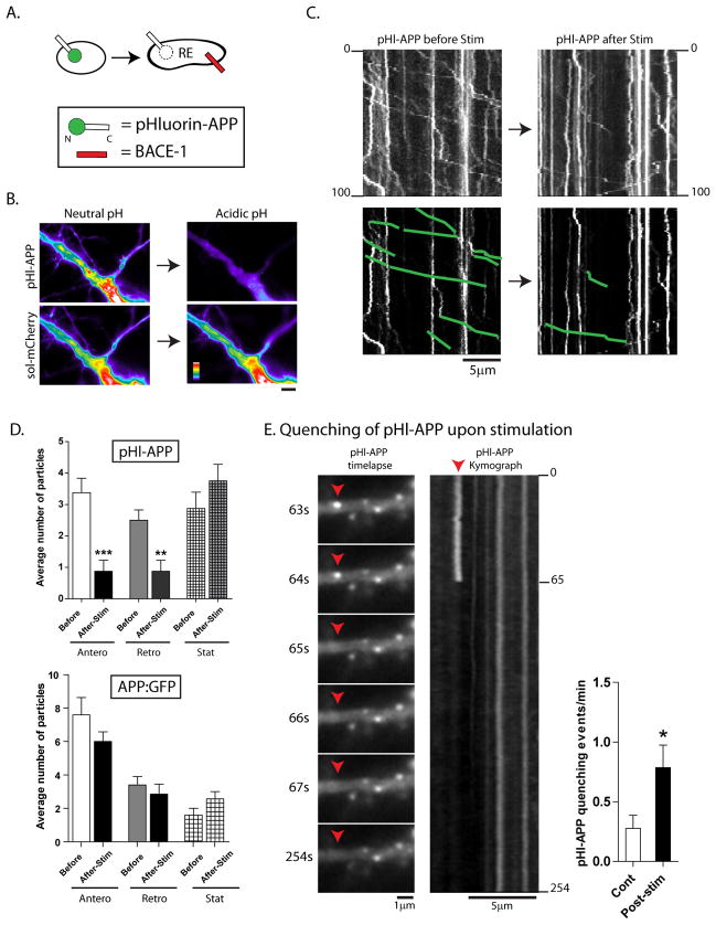
Figure 5. Mobile APP particles are routed into an acidic microenvironment upon activity-induction.
(A) Schematic of pHluorin:APP. Note that the pHluorin is tagged to the N-terminus (intra-luminal) end, and would be expected to quench if APP enters into vesicles with an acidic luminal pH (i.e., recycling endosomes).
(B) To test the pHl:APP construct, neurons were co-transfected with pHl:APP and soluble mRFP, and transferred to an acidic environment (see methods). Note that while the fluorescence of the pHl:APP is dramatically quenched, there is no change in the fluorescence of soluble mRFP (all images are identically scaled, intensity ‘heatmaps’ shown).
(C) Neurons were transfected with pHl:APP, and vesicle-motility was analyzed – in the same dendrite – before and after stimulation. As shown in the representative kymographs (motile vesicles are traced below), though there was no change in the intensities of stationary APP particles, the number of mobile APP particles in dendrites were dramatically reduced after stimulation.
(D) Top: Quantification of motile APP particles highlight the dramatic decease upon stimulation, with little change in stationary vesicles. Bottom: Similar experiments with APP tagged to conventional GFP does not show significant decreases in mobile particles upon stimulation. n ≈ 100–200 particles analysed for each condition, from at least two separate cultures. *p < 0.05; **p < 0.01; ***p < 0.001; two tailed, unpaired t-test.
(E) Upon stimulation, instances of pHl-APP quenching were also seen as shown in selected frames from a time-lapse (also see Supplementary Movie 2), further suggesting that APP was routed into an acidic compartment upon induction of neuronal activity. (*p < 0.05; two tailed, unpaired t-test).

