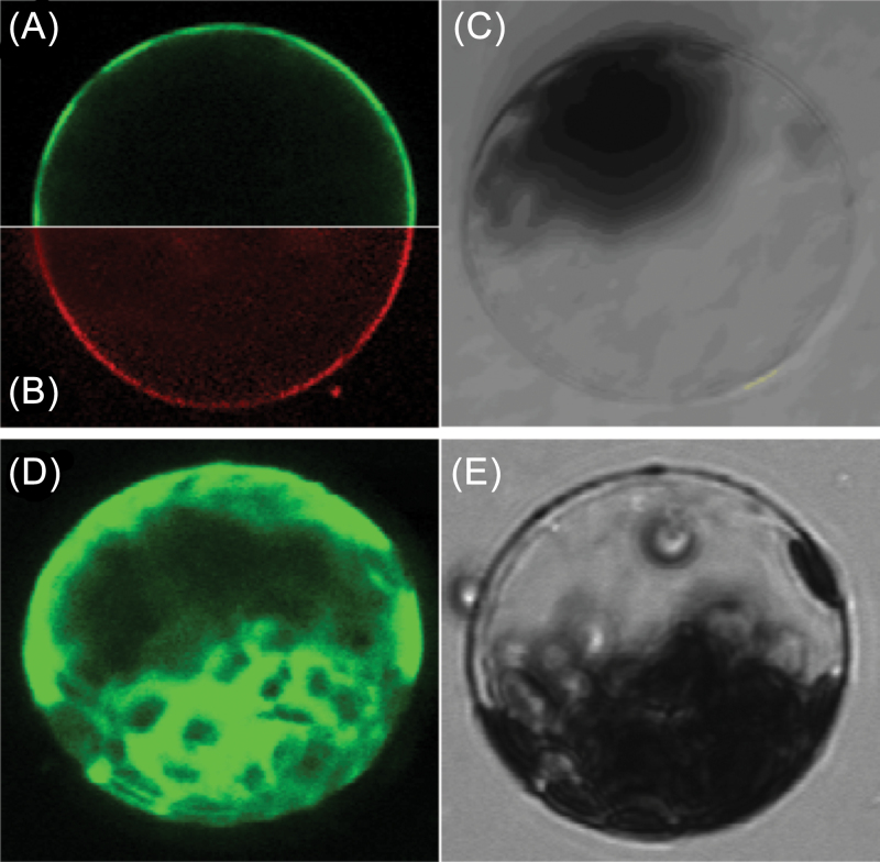
Fig. 4.
Subcellular localization of TaPI4KIIγ in Arabidopsis protoplasts. The TaPI4KIIγ–GFP fusion proteins (A) or GFP alone (D) driven by the 35S promoter were transiently expressed in Arabidopsis protoplast cells and observed under a laser scanning confocal microscope. In (B), the protoplast plasma membrane was labelled with the FM4-64 steryl dye. Bright field images are shown in (C) and (E). The scale bar is 15.38 μm. (This figure is available in colour at JXB online.)

