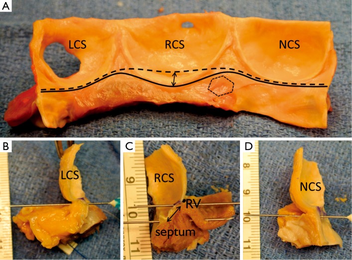
Figure 4.
A. Intraluminal view of longitudinally opened aortic root. Continuous black line delineates the ventriculo-aortic junction; the interrupted black line delineates the limit of proximal aortic root dissection; double black arrow show the segment of myocardium included into the base of the right coronary sinus; dotted line encircles the membranous septum (LCS, left coronary sinus; NCS, non coronary sinus; RCS, right coronary sinus); B. Longitudinal section through the nadir of the left coronary sinus; the limit of proximal root dissection reaches the level of the VAJ; C. Longitudinal section through the nadir of the right coronary sinus; the limit of proximal root dissection does not reach the level of the VAJ; double black arrow show the segment of myocardium included into the base of the right coronary sinus (RV, right ventricle; septum, interventricular muscular septum); D. Longitudinal section trough the nadir of the non-coronary sinus; the limit of proximal root dissection reach the level of the VAJ

