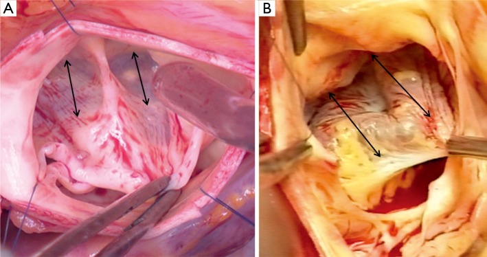
Figure 5.

Intraoperative pictures showing the so called ‘sinking sinus’, showing large segment of myocardium included into the base of the right coronary sinus. (A. Type 1 bicuspid aortic valve; B. Type 0 bicuspid aortic valve, double arrows show the width of the myocardial inclusion)
