Fig. 1.
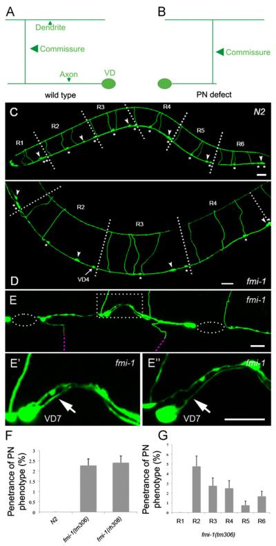
PN defects in fmi-1 animals. ((A) and (B)) A cartoon of a VD neuron in wt (A) and fmi-1 animals (B). (C) In juIs76 animals, the D-type GABAergic neurons can be organized in six regions (R1–R6) along the A/P axis. Regions R1–R5 each contain one DD (arrowheads) and two VDs (asterisks) whereas region R6 contains one DD and three VDs. VD and DD commissures are positioned anterior to their respective cell body. (D) In this fmi-1 animal, VD4 commissure is in region R3 instead of R2. As a result, R2 and R3 contain two and four commissures, respectively. In contrast, R2 and R3 in wt always contain three commissures as depicted (C). ((E)–(E’)) Ventral view of another fmi-1 animal where VD7 is displaying a PN (arrow). (E”) Rotated version of (E’). (F) Quantification of the PN phenotype penetrance. (G) Quantification of the PN phenotype per region. Dashed ellipse indicates regions lacking neuronal processes in the VNC whereas purple dashed lines show commissures out of focus. Dashed square show region magnified and depicted in ((E’)—(E”)). Scale bar is 20 μm in ((C) and (D)) and 10 μm in ((E)–(E”)). Anterior is to the left.

