Fig. 2.
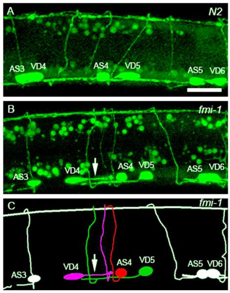
VD neurons display PN defects in fmi-1;lhIs35 animals. (A) L2 wt animal expressing the lhIs35 marker. VD neurons are posterior to AS neurons. (B) VD4 displaying a PN defect (arrow) in an L2 fmi-1 animal. In spite of this defect, VD4 still projects a ventrodorsal commissure. VD5 has a normal AN. Note that VD6 commissure is not shown in the fmi-1 animal. (C) A cartoon of (B). Scale bar is 10 μm.

