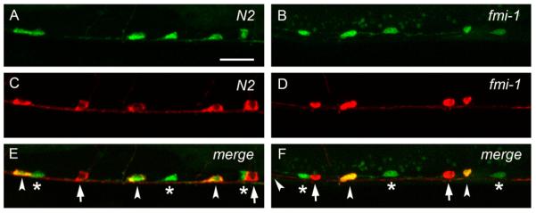
Fig. 3.

VD neurons are positioned posterior to AS neurons in wt and fmi-1 animals. ((A) and (B)) lhIs35 labels VD and AS neurons in wt (A) and fmi-1 (B). ((C) and (D)) AS neurons are labeled by RFP (Punc-53::tagRFP) in wt (C) and fmi-1 (D). ((E) and (F)) Merge views. VD neurons (asterisks) are located posterior to AS (arrowheads) in both backgrounds. Notice that the first AS neuron, from left to right, is not shown in (F). Red marker is also expressed in the DA neurons (arrows in E and F). Scale bar is 10 μm.
