
Figure 2. Prostate Hemorrhage following Biopsy Complicates Cancer Detection on T2-Weighted Imaging.
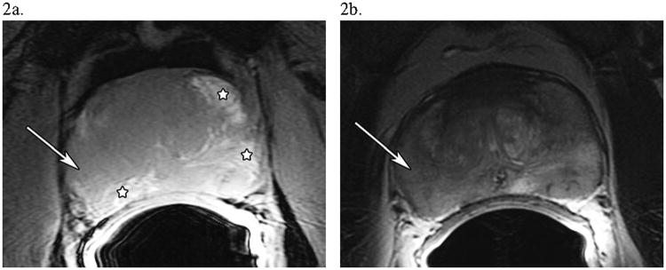
A 52 year-old man with PSA 5.8, Gleason 5+4=9, T2cN0M0 prostate cancer. (a) An axial T1-weighted image of the prostate showing possible areas of hemorrhage, which have a hyperintense signal (major hemorrhagic regions are starred). (b) An axial T2-weighted image of the prostate at the same level showing diffuse T2 signal hypointensity in the peripheral zone, indicating either tumor or hemorrhage. The white arrow in both (a) and (b) in the lateral region of the right side of the prostate shows an area suspicious for tumor, as there is T2-weighted signal hypointensity without T1-weighted signal hyperintensity.