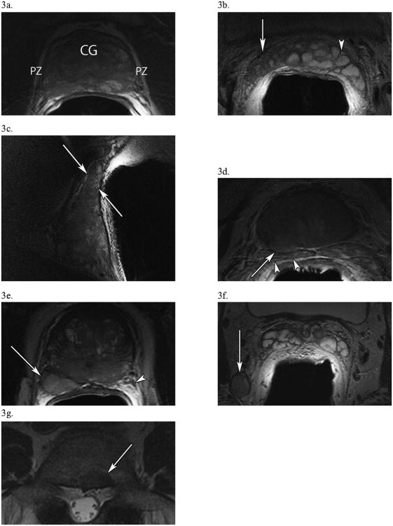
Figure 3. T2-Weighted Images of Critical Findings in Prostate Cancer.
(a) In a 61 year-old man with PSA 3.7, Gleason 3+3 =6, T2cN0M0 disease, an axial view of the prostate presenting a diffuse, capsule-contained peripheral zone (PZ) lesion with a “chalky” appearance. Notice how the PZ, usually hyperintense and clearly demarcated from the central gland (CG) which it surrounds, is now heterogeneous. (b, c) A different, 56 year-old man with PSA 9.3, Gleason 3+4=7, T3bN0M0 disease shows right-sided seminal vesicle invasion (white arrows) by tumor in axial (b) and sagittal (c) images. In the axial image (b), compare the hypointense, diseased right SV (white arrow) to the normal, hyperintense SV on the left (arrowhead). (d) In a 60 year-old man with PSA 134, Gleason 3+4=7, T4N0M0 disease, an axial image of the base of the prostate showing right-sided extracapsular extension (arrow) at 7 o'clock with probable involvement of the right SV (arrowheads). (e) In a 65 year-old man with PSA 40.1, Gleason 5+4=9, T4N1M1a disease, an axial view of the prostate displaying a large posterolateral right peripheral zone lesion (arrow) at 7 o'clock which invades the right neurovascular bundle. This lesion starkly contrasts with the normal NVB on the left (arrowhead). (f) From the same patient as (e), significant pelvic lymphadenopathy is present in an axial view at the level of the seminal vesicles, including this 1.5 × 1.3-cm, right posterior obturator node (arrow). (g) In a 75 year-old man with PSA 11.4, Gleason 4+4 = 8, T3aN0M1b prostate cancer, an axial view of the spine at L5 demonstrating a 1.5 × 1.1-cm metastatic bone lesion.

