Fig. 7.
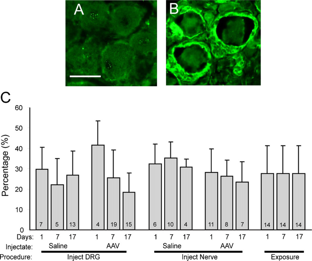
Identification of activated satellite glial cells in the dorsal root ganglion (DRG) as an indication of inflammation. (A) DRG tissue stained with anti-glial fibrillary acidic protein (GFAP) antibody 17 days after surgical exposure alone shows no surrounding rings of GFAP-positive sells. (B) DRG 17 days after AAV injection shows positive GFAP staining in rings of activated satellite glial cells surrounding neuronal somata. (C) Bars indicate the percentage of neuronal somata surrounded by GFAP-positive rings under different conditions, including different times after injections of saline or adeno-associated viral (AAV) vector, or exposure of the ganglia without injection. The numbers in the bars indicate the number of fields quantified, from two animals in each case. Mean ± SD. Statistical analysis (ANOVA) showed no differences between groups when different days were combined into a single group.

