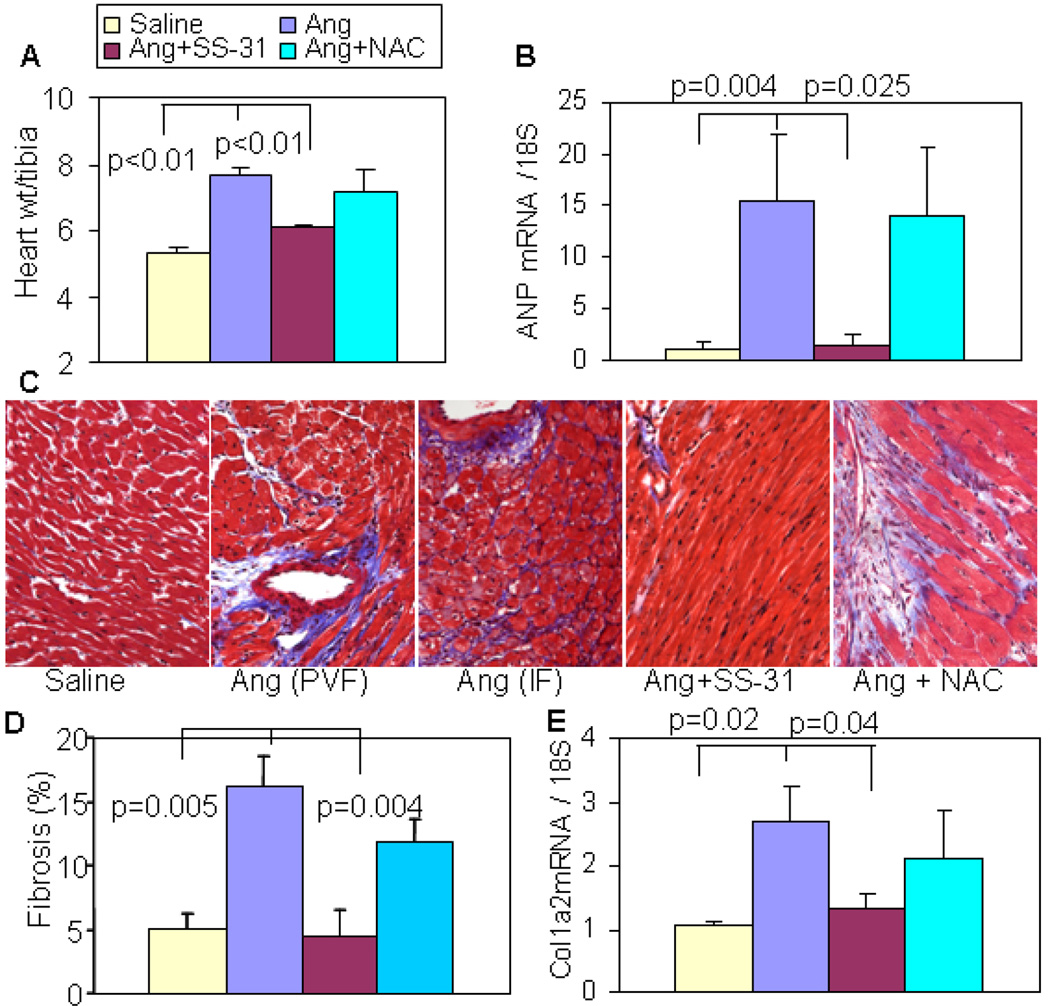
Figure 4. Ang-induced cardiac hypertrophy and fibrosis were attenuated by SS-31 but not oral NAC.
(A) Ang significantly increased heart weight (normalized to tibia length), and this was significantly attenuated by SS-31 but not by NAC. (B) qPCR showed a dramatic increase in ANP gene expression, which was significantly prevented by SS-31, but not by NAC. (C) Representative histopathology showing that Ang induced substantial perivascular fibrosis (PVF) and interstitial fibrosis (IF), which was protected by SS-31 but not NAC. (D) Analysis of blue trichrome staining demonstrated a significant increase in ventricular fibrosis after Ang, and this was substantially attenuated by SS-31 but not NAC. (E). qPCR showed upregulation of pro-collagen1a2 mRNA after Ang, which was significantly reduced in SS-31 hearts but not in NAC hearts. n=4–7.

