
Figure 2.
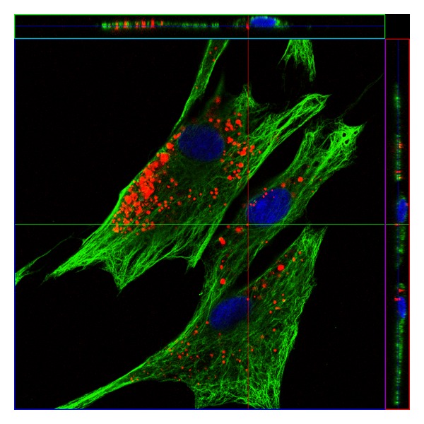
Spatial distribution of QDs (red) within the cells. Confocal image of labeled PSCs immunostained for Vimentin (green). Nuclei were stained with DAPI (blue).
Official websites use .gov
A
.gov website belongs to an official
government organization in the United States.
Secure .gov websites use HTTPS
A lock (
) or https:// means you've safely
connected to the .gov website. Share sensitive
information only on official, secure websites.
Spatial distribution of QDs (red) within the cells. Confocal image of labeled PSCs immunostained for Vimentin (green). Nuclei were stained with DAPI (blue).