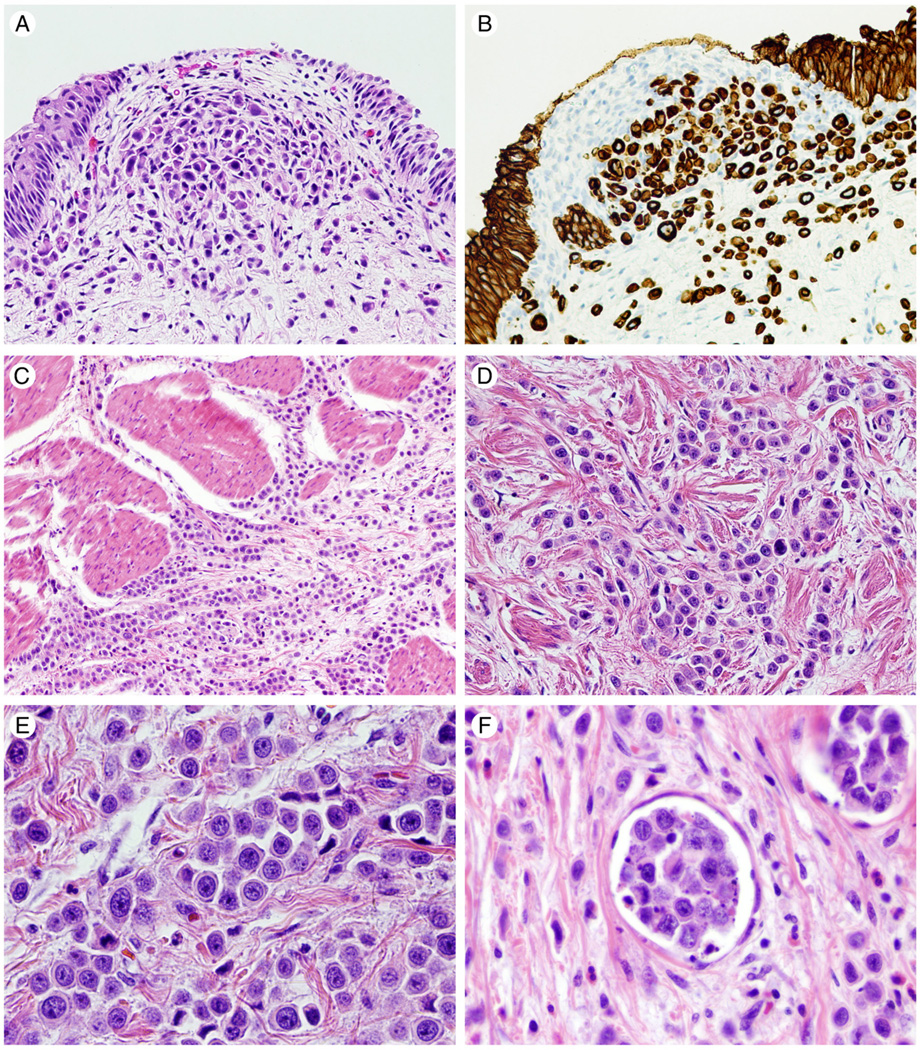
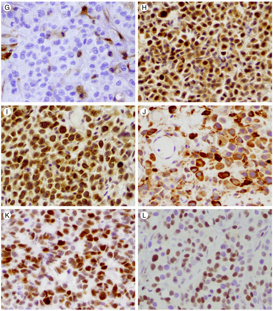
Fig. 1.
Hematoxylin and eosin stained photomicrographs of plasmacytoid urothelial carcinoma are shown in panels A, C, D, E, F. Muscularis propria invasion is shown in C and lymphovascular invasion in F. (×100 and ×400, respectively). B reveals plasmacytoid urothelial carcinoma immunohistochemical positivity for CK8/18 (×200). Representative immunohistochemical staining for PTEN, mTOR pathway members, c-myc, and p27 in tumor cells are illustrated in 1G-1L (×400). G illustrates loss of PTEN staining. Positive PTEN staining in endothelial cells is used as an internal control. H reveals nuclear and cytoplasmic staining for phos-AKT. Cytoplasmic staining for phos-mTOR and phos-S6 is shown in I and J respectively. K and L depict nuclear staining for c-myc and p27, respectively.


