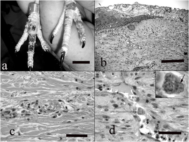
Figure 2.
a) Note numerous foci of dark discoloration and ulceration on the dorsal aspects of the feet, corresponding to vascular lesions detected histologically. Bar = 2 cm. b). Full thickness section of skin from the dorsal aspect of the toe, showing chronic lesions. Note ulceration, crust formation, epidermal hyperplasia adjacent to the ulcer, and dermal scarring. HE, bar = 300 μm. c) A venule in the dermis showing acute fibrinoid necrosis with thrombosis and mild perivascular inflammatory infiltrate. HE, bar = 150 μm. d) Small vein in the dermis showing endothelial cell hypertrophy, necrosis and exfoliation. Note intracytoplasmic schizont in a hypertrophied endothelial cell (arrow). HE, bar = bar = 60 μm. Inset) higher magnification of the infected endothelial cell. Note the numerous zoites fill the cytoplasm and displace the host nucleus (arrow). HE, bar = 10 μm.

