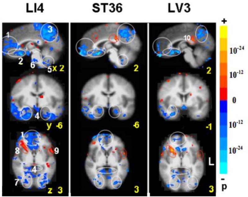
Fig. 1.
BOLD fMRI during acupuncture at 3 acupoints performed in randomized order (LI4 64 runs/37subjects, ST36 74/43, LV3 63/37). Clusters of deactivated regions appeared at the mid and ventral levels of the MPFC (1, 2), MPC (3) and MTL (4). Deactivation also occurred in the cerebellar tonsil and vermis (5), pontine nucleus (6) and extrastriate cortex (7). The general pattern was similar for all points with differences in magnitude of signal change and preferential localizations. Robust changes in all the regions were seen with LI4. Deactivation of the FP, pregC and cerebellum was minimal with LV3. A few paralimbic regions showed activation instead: the right anterior insula (8) and the postC-BA23d (9) with LI4 and LV3. The superior temporal gyrus BA22 (10) showed activation with all points. p< 0.0001

