Fig. 2.
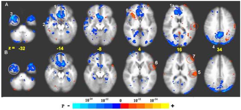
BOLD response: acupuncture vs. tactile stimulation at the right LI4, ST36, and LV3 acupoints performed on the same 37 subjects, p < 0.001. Deactivation network: In acupuncture (A) extensive deactivation appeared in the MPFC (1), MTL (2), TP (3) and MPC (4) regions. In tactile stimulation (B), deactivation appeared in similar regions but was more limited in extent than in acupuncture. The difference was most marked with the FP and pregC (1a) and PCN/BA31 (4). Activation network: Activation of SII (5), BA22 (6) and DLPFC (7) was more prominent in tactile stimulation than in acupuncture. The right anterior insula (8) notably showed strong activation with acupuncture but not with tactile stimulation (For details, see also Table 1).

