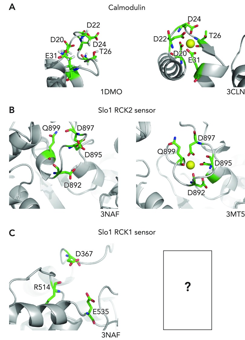
FIGURE 2.
Ca2+ coordination
Ca2+ coordination in calmodulin (A), the Slo1 RCK2 sensor (B), and the Slo1 RCK1 sensor (C). For each, the Ca2+-free apo structure is shown at left and the Ca2+-bound structure is shown at right. The image was rendered in MacPyMol version 0.99. The Ca2+-ligating residues are shown using sticks. Ca2+: yellow sphere; carbon: green; nitrogen: blue; oxygen: red. Water molecules participate in Ca2+ coordination, but they are not shown.

