
Fig. 8.
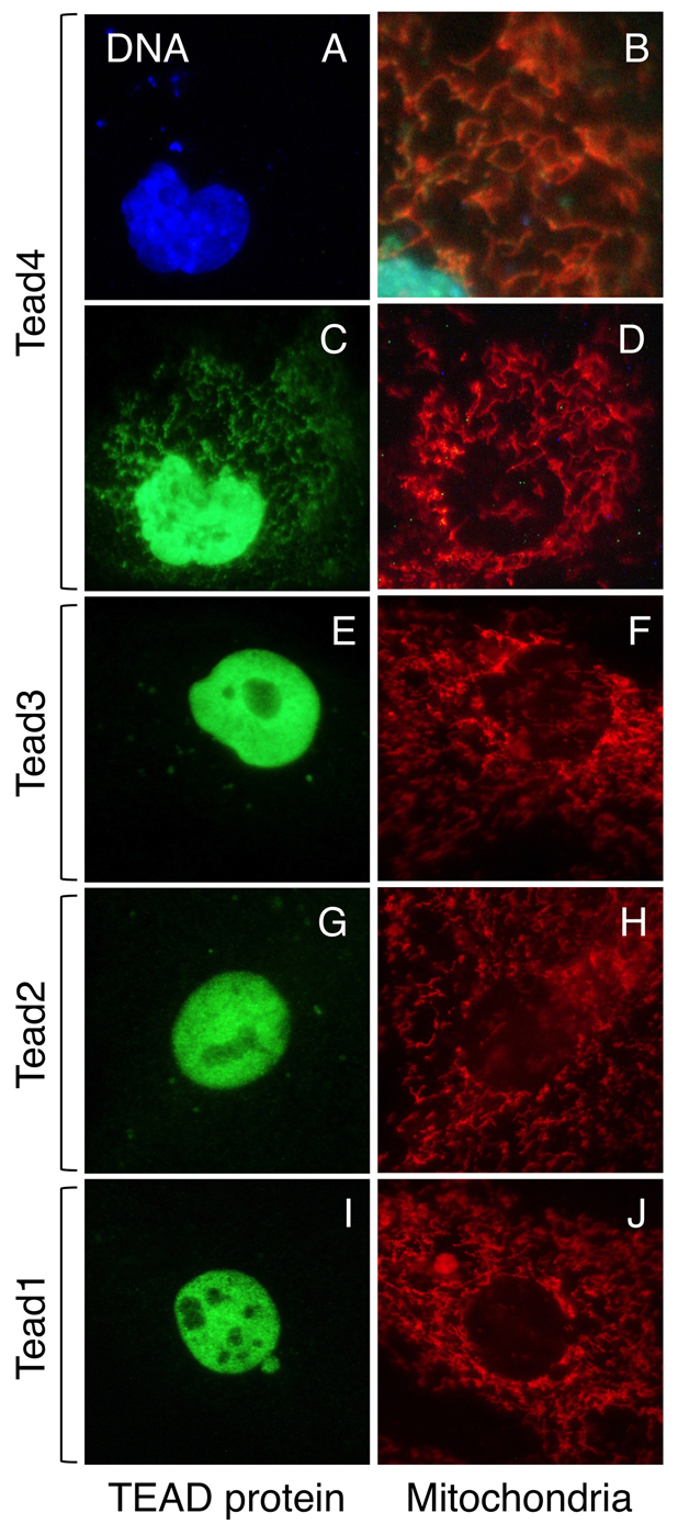
TEAD4 localizes to mitochondria as well as to nuclei, whereas TEAD1, TEAD2 and TEAD3 localize only to nuclei. (A-J) The indicated hemagglutinin epitope (HA)-tagged TEAD protein was expressed transiently in PMEFs. Forty-eight hours post-transfection, cells were stained with Mitotracker Red, then fixed and stained with anti-HA antibody (green). Cells were also stained with DAPI to visualize nuclear DNA (blue). TEAD4 (A-D) localized to both the nucleus and the mitochondria in same cell. Superimposition of C and D confirmed colocalization of TEAD4 (green) and mitochondria (red) that appear yellow in the enlarged section (B). By contrast, none of the cells transfected with TEAD1 (I,J), TEAD2 (G,H) or TEAD3 (E,F) showed mitochondrial localization of these HA-tagged proteins.