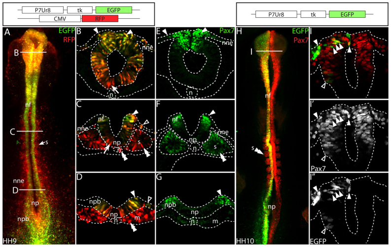
Fig. 2.
Enhancer P7Ur8 is co-expressed with Pax7 in the neural crest, but not in the somites. (A-D) Co-electroporation of red-marker control and P7Ur8-EGFP reveal specific expression at HH9 in the dorsal most part of the neural tube (B, closed arrowheads) and neural folds (C and D, closed arrowheads), but not in the ventral neural tube (B, arrow) or neural plate (C, arrow). P7Ur8 is expressed in some NNE adjacent to the neural folds (C and D, open arrowheads), but is not expressed in the somites, the notochord (C, double arrowheads) or the pre-somitic mesoderm (D, double arrowhead). (E-G) Sections showing normal Pax7 expression (green) in a similarly staged embryo, in the tips of the forming neural folds (F,G, closed arrowheads), in the dorsal neural tube and in migrating NC (E), and in the somites (F, double arrowhead). (H-I′) At HH10, P7Ur8 (green) is co-expressed with Pax7 protein (red). Cells in the dorsal neural tube strongly express P7Ur8 (I-I′, closed arrowheads), whereas weak P7Ur8 expression is seen in more ventral cells (I-I′, open arrowheads). n, notochord; m, mesoderm; np, neural plate; npb, neural plate border; nne, non-neural ectoderm; nf, neural folds; s, somites.

