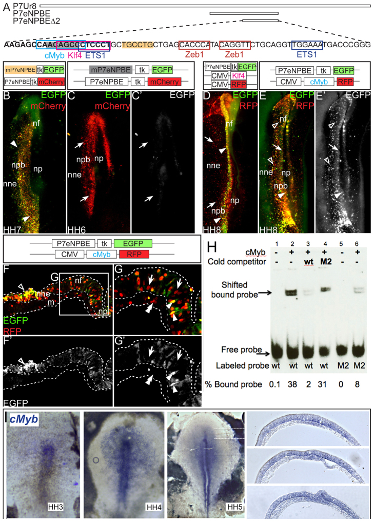
Fig. 4.
The transcription factor cMyb is crucial for the enhancer activity of P7eNPBE. (A) Position size and sequence of the enhancer P7eNPBEΔ2 in comparison with P7Ur8 and P7eNPBE. (B-C′) Putative DNA-binding sites of four selected TFs shown in colored boxes (cMyb, light blue; Klf4 pink; ETS1, dark blue; Zeb1, red). Two examples of mutated areas are indicated, with yellow for early unaltered (B, arrowheads) and gray for abrogated (C,C′, arrows) reporter expression. (D-G′) These TFs were electroporated to assess effect on P7eNPBE enhancer activity. (D) Overexpression of Klf4 (red) led to normal P7eNPBE-EGFP expression in neural folds and neural plate border (arrowheads) and to exclusion from the NNE (arrows). (E-G′) cMyb (red) overexpression led to reduced P7eNPBE-EGFP signal in the neural folds (E-E′, arrow) with residual salt and pepper patterns, and expansion in the NNE (E,E′, open arrowheads). (F-G′) Sections of a similarly treated and staged embryo showing P7eNPBE expression in the NNE (open arrowheads, F,F′) and neural plate (arrowheads, G). P7eNPBE-EGFP is not ectopically activated in the mesoderm (double arrowheads G) and is much reduced in the neural folds, but displays a ‘salt and pepper’ pattern, with some cells lacking (arrows), and others presenting, expression (arrowheads) (compare with Fig. 2C,D). (H) EMSA performed using a 20 bp probe (bold sequence in A) containing the wild-type cMyb-binding site or the M2 mutated version. When cMyb is added, it binds to the wild-type probe, generating a clear shift (lane 2 ∼38% bound probe), which is severely reduced when cold unlabeled wild-type probe is added (lane 3, ∼2%); instead, adding the same excess amount of cold unlabeled mutated M2 probe still displays a robust shift (lane 4, ∼31%); however, only 8% of the labeled mutated-M2 probe is bound by cMyb (lane 6). (I) cMyb is expressed in the early ectoderm of chick embryos prior to Pax7. Whole mount in situ hybridization on HH3-5 chick embryos. Dotted lines at HH5 indicate sections shown on the left. m, mesoderm; np, neural plate; npb, neural plate border; nne, non-neural ectoderm; nf, neural folds.

