Figure 1.
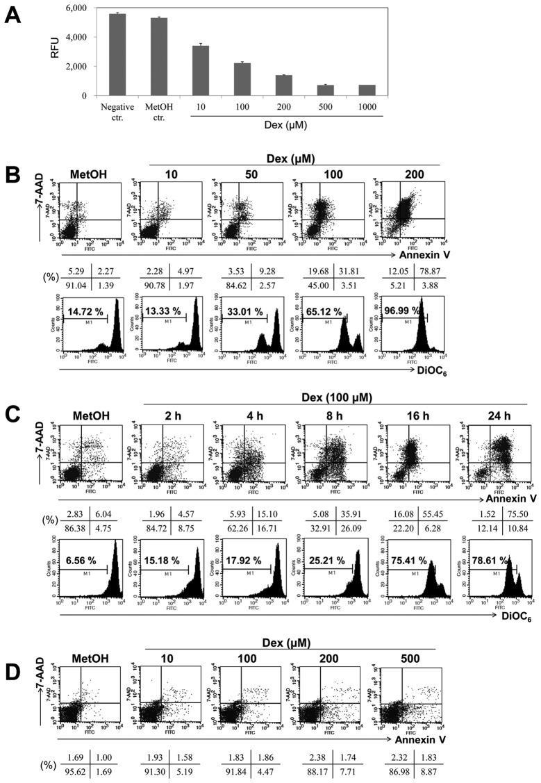
Dex induced apoptosis in a dose- and time-dependent manner in EBV-transformed B cells. (A) EBV-transformed B cells (5×10⁴ cells/well) were cultured in 96-well plates. After 24 h, cell proliferation was measured by AlamarBlue assay. RFU is the relative fluorescence unit. (B and C) EBV-transformed B cells and (D) PBMCs were treated with 10, 50, 100 and 200 μM of Dex for 2, 4, 8, 16 and 24 h. The percentage of apoptotic cells was estimated by Annexin V/7-AAD staining. Dot plot graphs show percentage of viable cells (Annexin V−/7-AAD−), early-phage apoptotic cells (Annexin-V+/7-AAD−), late-phage apoptotic cells (Annexin-V+/7-AAD+), and necrotic cells (Annexin-V−/7-AAD+). To measure disruption of Δψm, cells were stained with DiOC6. Diminished DiOC6 fluorescence indicates Δψm disruption and percentages indicates the cell proportion in each bar. Results are representative of three independent experiments.

