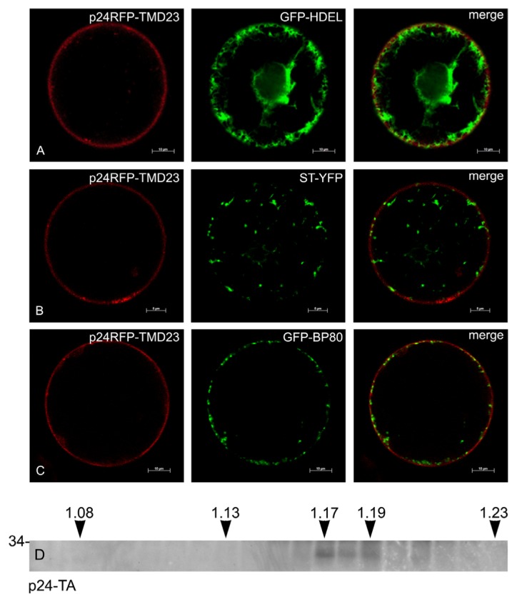
Figure 10.
Subcellular localization of p24RFP-TMD23 and p24-TA. A, B and C: p24RFP-TMD23 was transiently co-expressed in tobacco protoplasts for 24 h with markers for the ER (GFP-HDEL), trans-Golgi cisternae (ST-YFP) or TGN/MVB (GFP-BP80). Scale bars: 10 μm (A and C); 5 μm (B). (D) Leaves of transgenic tobacco expressing p24-TA were homogenized in the absence of detergent. Subcellular compartments were separated by ultracentrifugation on isopycnic sucrose gradient. Proteins in each fraction were finally analyzed by SDS-PAGE and protein blot with antibodies against p24. Top of the gradient is at left; numbers on top indicate fraction density (g/mL). The number at left indicates the position of molecular mass marker, in kDa.

