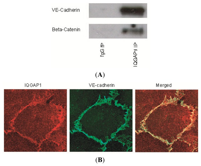
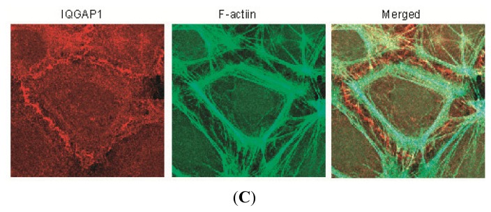
Figure 1.
IQGAP1 associates with vascular endothelial (VE)-cadherin, β-catenins and F-actin. (A) HUVEC cell lysate was immunoprecipited for IQGAP1, VE-cadherin and β-catenin. Data represent three independent experimental determinations; (B) HUVECs were also grown to confluence on precoated glass coverslips and stained for IQGAP1 and VE-cadherin. Primary antibodies were visualized with Alexa Fluor 488-labeled goat anti rabbit IgG (green) and Alexa Fluor 594-labeled goat anti mouse IgG (red). Yellow indicates where colocalization occurs. IQGAP1 was shown to colocalize with VE-cadherin at cell-cell contact sites; (C) IQGAP1 was visualized at the intercellular junctions (Red) and did not colocalize with F-actin (Green, Phalloidin-488).


