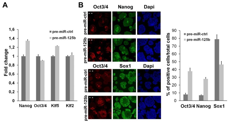
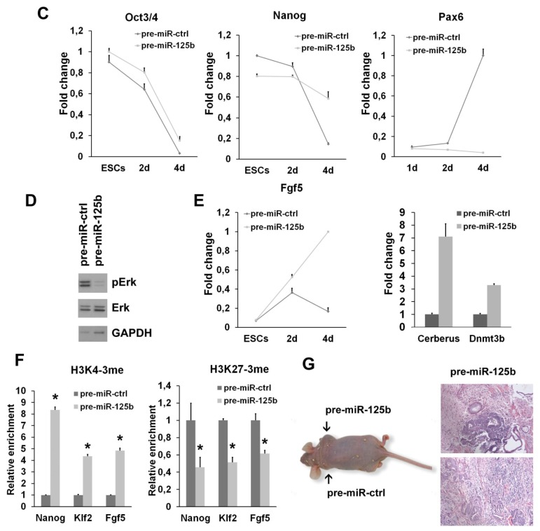
Figure 2.
Effects of miR-125 ectopic expression in vitro and in vivo. (A) Analysis of stemness markers (Oct3/4, Nanog, Klf2 and Klf5) in undifferentiated ESCs transfected with pre-miR-125b or with control pre-miR (pre-miR-ctrl). The fold change is calculated by assigning the arbitrary value, one, to the amount found in cells transfected with control pre-miRNA; (B) Immunofluorescence analysis of four-day differentiated SFEBs upon miR-125b overexpression. Markers of pluripotency (Oct3/4, Nanog) and neuroectoderm (Sox1) are shown. Scale bar: 50 μm; (C) qPCR analysis of stemness (Oct3/4 and Nanog) and neuroectodermal (Pax6) markers in differentiating ESCs upon miR-125b overexpression. The fold change is calculated by assigning the arbitrary value, one, to the time point showing the highest amount of the indicated mRNA; (D) The level of active ERK (P-ERK) were analyzed by Western blot in cells transfected with pre-miR-125b or the control pre-miR after four days of differentiation—the experiment shown in the Figure is representative of two independent experiments; (E) The level of the epiblast marker, Fgf5, was measured by qPCR in undifferentiated ESCs and during differentiation upon pre-miR transfection. The epiblast markers, Cerberus and Dnmt3b, were measured at four days of SFEB differentiation in the cells transfected with the indicated pre-miR. The fold change is calculated as indicated in (C); (F) ChIP-qPCR analysis was performed on chromatin from ESCs transfected with the indicated pre-miR and induced differentiation for four days as SFEBs. The graphs show the methylation state of histone H3 on the promoters of pluripotency (Nanog and Klf2) and epiblast (Fgf5) markers. Data are expressed as fold enrichment relative to the control; (G) Immunodeficient mice were injected with ESCs transfected with the pre-miR-125b (right side) and ctrl pre-miR (left side) after three days of differentiation in vitro (left panel). Teratomas generated by ESCs overexpressing miR-125b were explanted after one month, and the tissues were analyzed after eosin-hematoxylin staining (right panels) (*p < 0.05).


