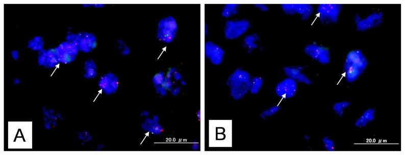
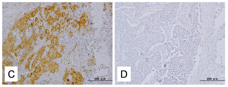
Figure 1.
Dual-color fluorescence in situ hybridization (FISH) validates amplification of the KRAS or MAPK1 gene in type II ovarian carcinoma. (A) FISH analysis of KRAS showed a homogeneously stained region in a tumor with gene amplification. White arrows indicated amplification of KRAS gene; (B) FISH analysis of MAPK1 showed a homogeneously stained region in a tumor with gene amplification. White arrows indicated amplification of MAPK1 gene; (C) Intense immunoreactivity toward p-ERK was present in both the nuclei and cytoplasm of carcinoma cells; (D) This sample shows negative case of staining for p-ERK.


