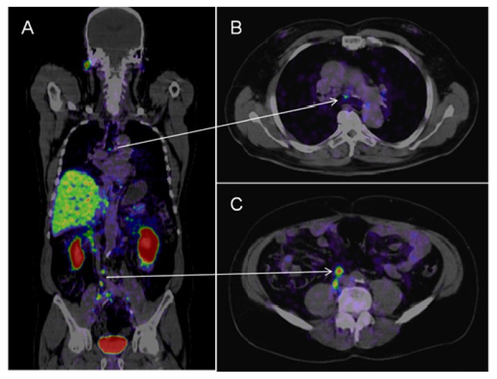
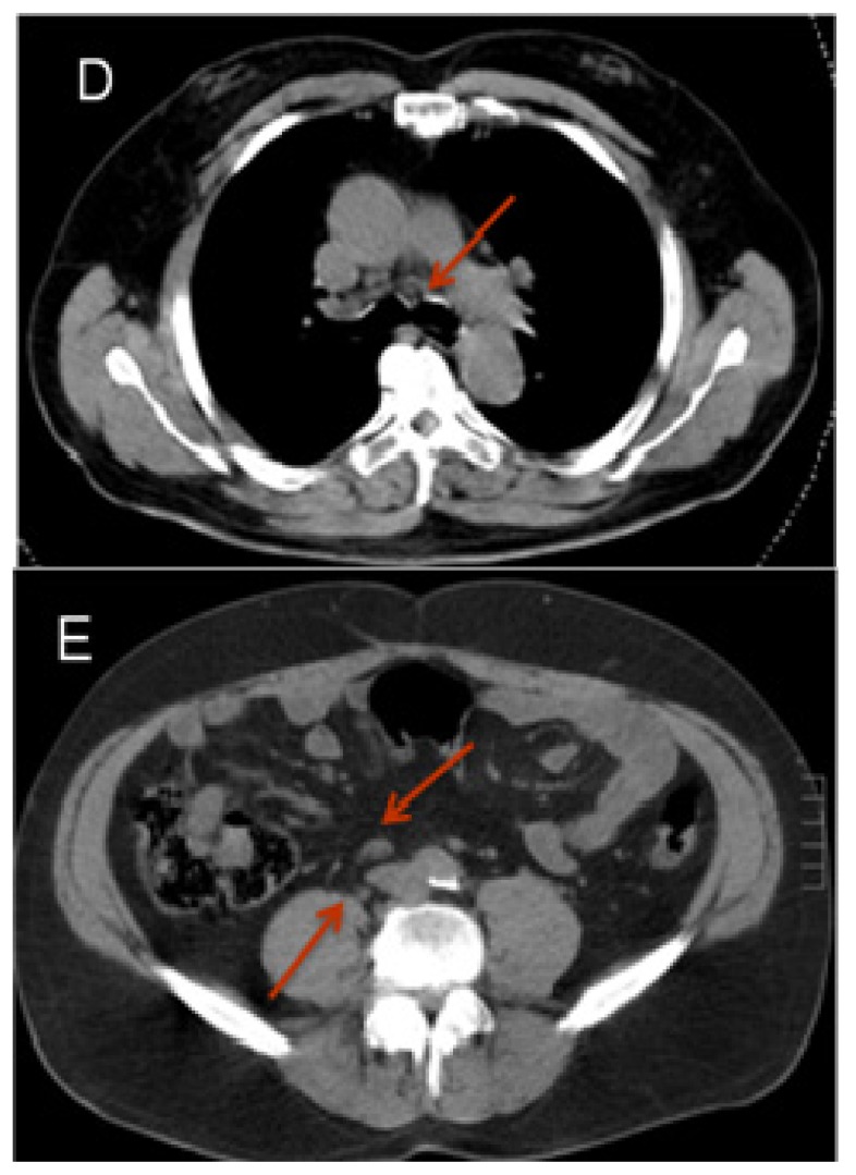
Figure 2.
68Ga-PSMA-PET-CT. Patient with prostate cancer (status after brachy-therapy and bilateral iliac lymph node dissection, current PSA 21 μg/L). PET images were acquired after the administration of 68GA-PSMA-Ligand (60 min thereafter). The figure shows fused images (PET-CT): On the coronal view (A) a pathologic isotope uptake in multiple lymph nodes in the right para-iliac region and infra-carinal (mediastinum) is clearly visible; the corresponding transversal images show the para-iliac (B) and infra-carinal (C) lymph nodes with elevated uptake of the tracer. The corresponding transversal native CT images present these suspect structures (D,E) as normal sized lymph nodes (marked by red arrows).


