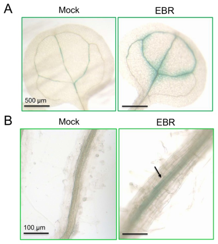
Figure 6.
Histochemical GUS staining of miR395a expression in A. thaliana. Expression patterns of miR395a promoter::GUS plants in (A) leaf and (B) root tissue. After growing for 7 days, seedlings were grown under EBR treatment (10 nM EBR) or mock control for 3 h. The arrow indicates a high concentration of miR395a distributed in the vascular bundle compared with the mock treatment.

