Figure 2.
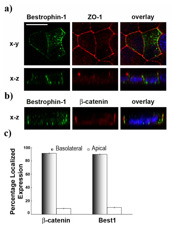
(a) Transfected MDCK cells express Best1 (green) at the basolateral surface, colocalizing with the tight-junction marker ZO-1 (red) on X-Y and X-Z single confocal scans; (b) Best1 (green) at the basolateral surface, colocalizing with the basolateral marker β-catenin (red) on X-Z single scan. Nuclei are in blue, scale bar = 10 μm; (c) Best1 localizes to the basolateral surface at the same level as the well-characterized marker β-catenin. Pixel intensity values obtained from confocal images per Z-focal plane of ten cells transfected with wild type BEST1 cDNA and the basolateral threshold in each cell were used to calculate percentage localized expression (mean ± SEM., n = 10, p = 0.018).

