Figure 4.
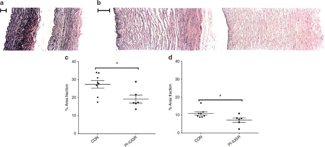
Verhoeff-van Gieson (VVG) histology and analysis. VVG-stained cross-sections for representative (a) carotid arteries (left side: control panel and right side: placental insufficiency–induced intrauterine growth restriction (PI-IUGR) panel; bar = 100 µm) and (b) umbilical arteries (left side: control panel and right side: PI-IUGR panel; bar = 200 µm). Elastin area fraction measures the collagen-to-elastin ratio. The (c) carotid and (d) umbilical arteries showed reduced area of elastin to total arterial wall area (*P < 0.05), demonstrating reorganization of collagen and elastin.

