
Fig. 1.
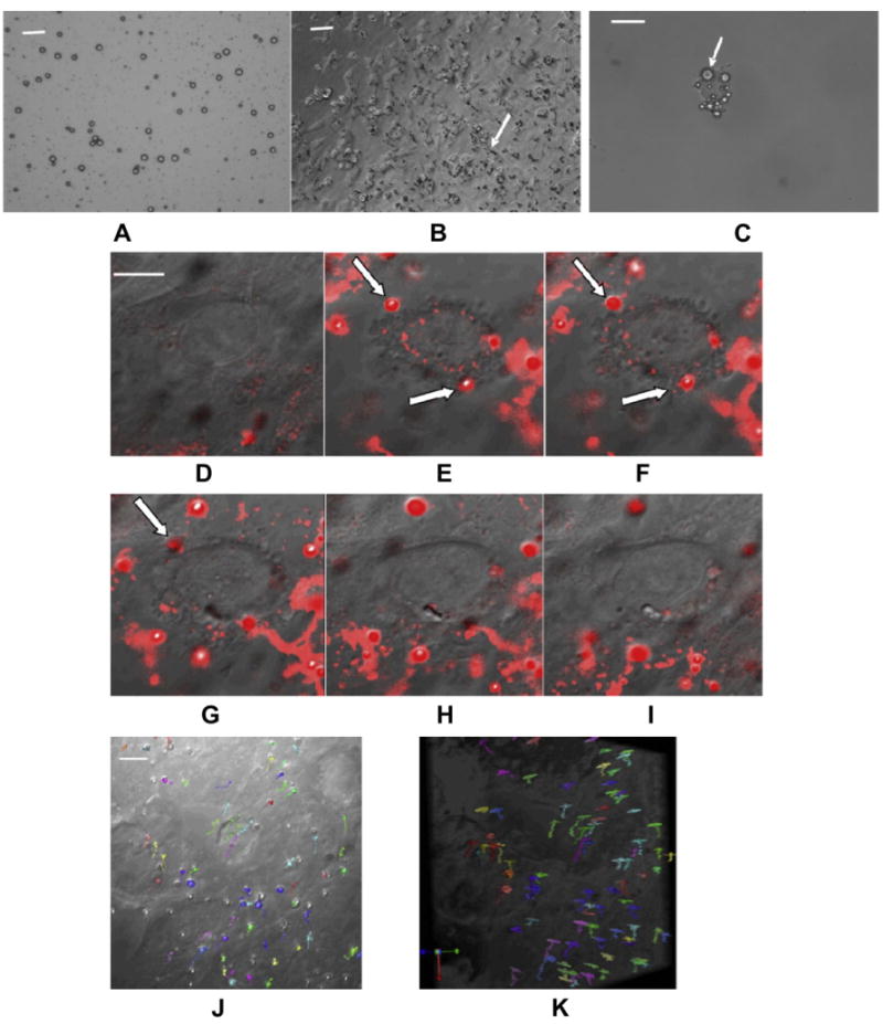
Labeling NPCs with cationic perfluorocarbon gas-filled MBs and tracking single bubbles inside NPCs. (A) DIC image of MB suspension (Scale bar = 10 μm). MBs are dark circles due to light diffraction; (B) DIC image of NPCs labeled with MBs (Scale bar = 20 μm); (C) A single trypsinized NPC containing multiple MBs (arrows) (Scale bar = 10 μm); (D–I) Time course showing the internalization of several MBs by NPCs (arrow) that disappear as they move out of the 0.8 μm slice (Scale bar = 10 μm); (J, K) On-end (J) and side view (K) of the 3-D stack acquired over an 8.8 μm thick slab shows the movement of MBs (each colored arrow) within the volume without localizing to a specific location (Scale bar = 10 μm).