
Fig. 2.
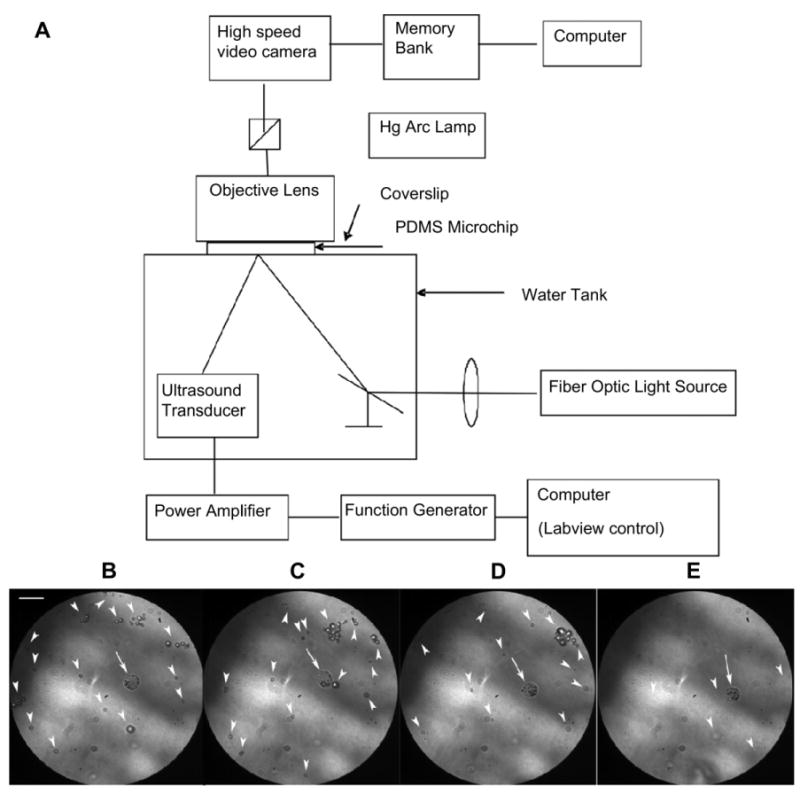
Optical observation of a MB-labeled NPC during insonation at 2.25 MHz and 1.5 MPa peak negative pressure. (A) Schematic of the experimental setup. Note that the focal points of the ultrasound pulse, the microscope, and the light were identical; (B–E) Representative frames from the high speed camera before (B) and following the 1st, 2nd, and 4th consecutive US pulses. Note that MBs within the NPC (arrow) remained intact after the 4th pulse while the many free MBs (arrowheads) diminished in number (E). All free MBs (arrowheads) were recognized from artifacts based on the video recording (See Supplement video) (Scale bar = 10 μm).