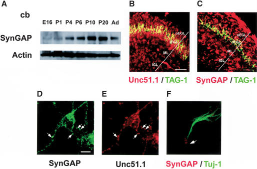
Figure 2.
Unc51.1 and SynGAP expression in developing cerebellum. (A) SynGAP protein is expressed throughout cerebellar development. Protein extracts prepared from varying stages of developing cerebellum (E16, P1, P4, P6, P10, P20, and adult) were analyzed by immunoblot. (B,C) Cellular distribution of Unc51.1 (B) and SynGAP (C) revealed by the antibody staining over P6 cerebellar sagittal sections (60 μm). TAG-1 antibody was used to mark differentiating granule cells in deeper external granular layer (iEGL). (oEGL) Outer external germinal layer that contains proliferating granule cells; (ML) molecular layer; (IGL) internal granular layer. (D,E) Subcellular distribution of SynGAP (D) and Unc51.1 (E) in granule neurons in culture. Arrows indicate punctae double-positive for Unc51.1 and SynGAP. (F) SynGAP localization in a growth cone (arrow). Tuj-1 was used to stain granule cell axons. Bar: D–F, 5 μm.

