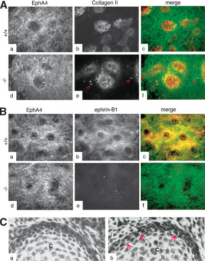
Figure 3.
Loss of ephrin-B1 does not affect the chondrogenic capacity of limb bud cells in vitro. (A) High-density micromass cultures from limb bud cells isolated from wild-type (panels a–c) or ephrin-B1 null (panels d–f) E11.5 embryos were analyzed by immunofluorescence using the EphA4 antibody (green) and a collagen II-specific antibody (red), as indicated. Collagen II-positive cells (red arrows) can be detected outside the chondrogenic micromasses in the culture isolated from mutant embryos. (B) High-density micromass cultures from limb bud cells isolated from wild-type (panels a–c) or ephrin-B1 null (panels d–f) E11.5 embryos were analyzed by immunofluorescence using the EphA4 antibody (green) and a monoclonal antibody specific for ephrin-B1 (25H11; red). Both EphA4 and ephrin-B1 are expressed in the cells surrounding the chondrogenic micromasses, including the perichondrium. (C) H&E staining of sections from wild-type (panel a) or ephrin-B1 heterozygous (panel b) E15.5 embryos. Space between cells can clearly be seen in mutant (arrows) but not in wild-type perichondrium. (Panel c) Chondrocytes.

