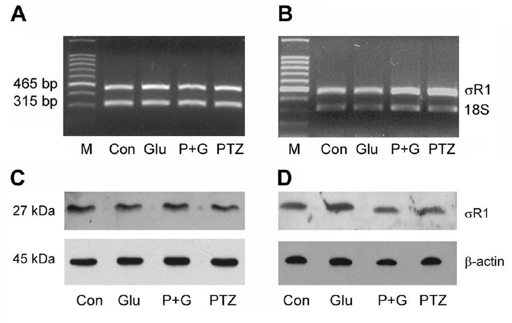
Fig. 5. σR1 mRNA and protein levels in 1°GC following treatment with Glu and (+)-PTZ.
1°GCs were incubated for 6 h (A & C) or 18 h (B & D) in the absence or presence of 25 µM Glu and the absence or presence of 3 µM (+)-PTZ. A and B: total RNA was isolated and used for semiquantitative RT-PCR. Primer pairs specific for mouse σR1 mRNA (465 bp) were used. 18S RNA (315 bp) was analyzed in the same RNA samples as the internal control. RT-PCR products were run on a gel and stained with ethidium bromide. C and D: Proteins were extracted from cells and subjected to SDS-PAGE, followed by immunoblotting using an affinity purified antibody against σR1, Mr ~27 kDa or β-actin, Mr ~45 kDa (internal loading control). (M, DNA marker; Con, control; Glu, 25 µM glutamate-treated cells; P + G, (+)-PTZ 3 µM plus 25 µM glutamate; (+)-PTZ, 3 µM (+)-PTZ incubation alone).

