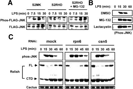
Figure 5.
Involvement of proteasomal degradation in Relish-dependent JNK inhibition. (A) S2MK and S2RHD cells were transfected with Flag-DJNK expression plasmid and then treated with CuSO4. S2RHD cells were untreated or further treated with 50 μM MG-132 for 2 h. After stimulating with LPS, cell lysates were prepared and analyzed by immunoblotting with antibodies specific to phos-JNK (upper panels) and Flag (lower panels). (B) S2 cells were treated with DMSO, 50 μM MG-132, or 5 μM lactacystin for 2 h and then unstimulated or stimulated with LPS for the length of time indicated. Phos-JNK was analyzed by immunoblotting. (C) S2 cells were treated with dsRNAs specific to rpn6 or csn5 for 3 d and then unstimulated or stimulated with LPS for the length of time indicated. Cell lysates were prepared and analyzed by immunoblotting with the antibodies indicated on the left.

